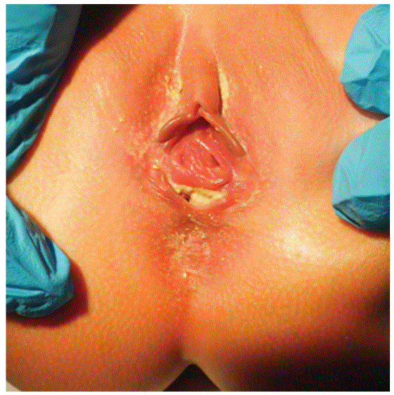
Figure 1: Visualization of Rectovestibular fistula.

Abiezer Disla* Dulce Gonzalez Wallis Tavarez George Vermenton
Department of Pediatrics, Lincoln Medical and Mental Health Center, Affiliated with Weill Cornell Medical College, New York, USA*Corresponding author: Abiezer Disla, Department of Pediatrics, Lincoln Medical and Mental Health Center, Affiliated with Weill Cornell Medical College, New York, USA, E-mail: abiezerd20@gmail.com
An imperforate anus is usually diagnosed at birth or shortly thereafter with physical examination being the cornerstone of diagnosis and guiding further management [1]. Despite routine postpartum physical examination, one in five neonates born with imperforate anus has delayed diagnosis [2]. Such a delay in diagnosis may complicate the surgical repair and may contribute to both functional and psychological problems for the patient and family. Constipation is one of the leading and most common symptoms of presentation [2]. In girls, due to a higher frequency of low anorectal malformation, lesions likely tend to be missed in neonates [3].
We describe an unusual case of an 18 month old female with chronic constipation due to delayed diagnosed of the imperforate anus with recto vestibular fistula in the absence of associated syndromes. Even though this condition in common, there have only been a few cases of the imperforate anus with enter vestibular fistula in the literature, presenting at this age in developed countries.
Our patient is an 18 Month old Hispanic girl, born full term appropriate gestational age by normal spontaneous vaginal delivery in the United States; the mother had standard prenatal care and an uncomplicated pregnancy. She underwent routine prenatal ultrasonography there was no reported evidence of congenital anomalies. The patient presented to Lincoln Hospital Emergency Department (ED), with a chief complaint of abdominal distention, vomiting, and history of chronic constipation.
She had bowel movements every 4-5 days described as small amount with straining during defecation. She also had abdominal distention associated with intermittent vomiting described as small in amount, with food contents, non-bloody and non-bilious. The patient had multiple visits to different emergency departments, and the mother was using Polyethylene glycol 3350 (Miralax), 1-2 times/ week, with a mild resolution of symptoms.
On physical examination, she was noted to have abdominal distention, normal bowel sound auscultated in all quadrants without organomegaly, normal external female genitalia, but no anal opening, instead a small opening was noted in the posterior aspect of the vaginal vestibule (rectovaginal fistula, Figure 1). Her exam was also remarkable for polydactyl of the left fifth digit. Her neurological exam was normal, as was the rest of her physical examination.
Figure 1: Visualization of Rectovestibular fistula.
Patient’s initial laboratory investigation were all within normal limits. She was referred to the pediatric surgeons for a further evaluation. Further studies did not show any other anomalies known to be associated with imperforate anus. Subsequently, patient was evaluated by a multidisciplinary team including general surgery and plastic surgery. A colostomy was performed followed by two more reconstruction surgeries. She has since recovered without any complications and is now doing well, awaiting for her final surgical procedure.
Imperforate anus, or anal atresia, is one of the conditions in the spectrum of anorectal malformations (ARM). It is known that this occurs during the 8th week of fetal gestation when the hindgut fails to completely develop. The normal and abnormal development of the hindgut is still a matter of speculation. However, as a result, recent studies most embryologic events that finally lead to abnormal hindgut development are better known than in the past:
It is not a rare entity; it occurs every 1 to 5,000 live births. The exact cause of an ARM remains unknown [5].
More commonly this malformation may occur as an opening in the urethra, bladder or vagina or as a blind pouch. Most commonly in females is the recto vestibular fistula, a communication between the rectum and the vaginal vestibule rarely is there a fistula between the rectum and vagina [5]. The presence of any type of fistula in the female can be misleading because meconium is passed and it is not always appreciated that the meconium is coming from an abnormal opening. Diagnosis depends mainly on a careful inspection of the perineum and on an insistence in seeing the orifice from which the meconium is being discharged [3]..
Late presentations in female’s anorectal malformations are quite common especially in low malformations and in third world countries where health care is limited. Unusual cases were reported by Rawat J, Singh S, Pant N, in a retrospective study conducted at King George medical University, in Lucknow, India. Study shows that out of 627 cases of ARM managed over the 5 years, ten girls (5.3%) presented between 12 and 18 years (average 14.4 years), and the main reasons for the delay in the presentation were misinformation, illiteracy, and poverty [6].
A studied published by Sinha SK, Kanoja RP, Wakhlu A, a review of data, from 2003-2006 done at King George medical University, in Lucknow, India, revealed, 43 patient included in the study, twenty one of these patients were male and twenty two females, varying from 7 days to 19 years. They found that fifteen of the girls that had low type ARMs all were passing stool from abnormal proximal opening. Eleven were born at home with no obstetrics care; out of all girls diagnosed only three cases presented with recto vestibular fistula. Their mode of delayed presentation differentiated from a developed country where constipation and abnormal anal opening detected by parents are more common. Their data analysis noted wrong advice regarding the correct age of treatment is the most common cause. The second most important cause was the inadequate management of the ARM elsewhere. Social factors were also a significant determinant owing to lack of financial and social support [7].
Kim, Gow, et al. [2] conducted a systematic reviewing analyzing data from 1987-1997 at the British Columbia’s Children’s Hospital, in the province of British Columbia, Canada. It revealed that a total of 13 patients with an ARM between 3 and 11 months of age because of increasing constipation. From the 13 patient only 1 patient presented at age of 11 months associated with recto vestibular fistula and no other associated anomaly [2]. Given the socio-economic similarities between the US and Canada the results of this study are similar to that of our case report.
Patients with an imperforate anus have an increased risk of having another congenital anomaly, most likely from the urogenital system, up to 40-45% [8]. Our patient went further evaluation, excluding vacterl defined by the presence of at least 3 of the following congenital malformations: vertebral defects, anal atresia, cardiac defects, tracheoesophageal fistula, renal anomalies and limb abnormalities.
A diagnosis of anal atresia is performed by a complete physical exam, therefore, in the majority of the time, the diagnosis is made after birth. A complete physical examination should include evaluating the anus and the perineum [8]. More than 90% of the time, the diagnosis in females can be established on perineal inspection [9].
Management of a patient found to have an imperforate anus includes an abdominal and pelvic ultrasound as well as radiographs of the spine and sacrum to rule out common coexisting conditions. Patients with this condition require an early and prompt diagnosis due to its increased mortality if left unrepaired. Some studies have reported a mortality rate of up to 30% for imperforate anus’ left unrepaired [3]. Complications range from abdominal distention and colonic perforation to sepsis and electrolyte imbalance.
A diagnosis is considered delayed if it’s made 48 hours after the birth of the neonate. A retrospective study published by Turowski, C, in 2010, in patients with imperforate anus, show 21.2% of their patient had delayed diagnosis more than 48 hours after birth [1].
Once the diagnosis of anal atresia is made, patients are referred urgently to the surgeon. Surgical management depends on the type or malformation and expertise of the surgeon, it can be done in a single intervention or multiple steps operations. The usual timeline for the surgeries are, colostomy at birth, followed by definitive surgery at 2-3 months of age followed by closure of the colostomy at 6 months old [3]. The main goal of surgical repair is for the patients to achieve a good quality of life. Fortunately, the majority of the patients with an ARM who undergo repair have good bowel function. The patients with more moderate to severe anomalies may have occasional fecal soiling. 8 The best approach for these patients is to provide an adequate bowel regimen with the use of enemas, colonic irrigations or suppositories [8].
Comprehensive physical examination including thorough evaluation of the recto-anal region, physiological function, together with a complete detailed history remained the cornerstone for identification of constipation in children. Precise diagnosis of imperforate anus, including a proper rectal exam, is essential to guide appropriate interventions, prevent complications and avoid significant life-threatening morbidity.
Download Provisional PDF Here
Article Type: CASE REPORT
Citation: Disla A, Gonzalez D, Tavarez W, Vermenton G (2018) Chronic Constipation due to Undiagnosed Imperforate Anus with Rectovestibular Fistula in an 18 Month Old Female. J Clin Case Stu 3(3): dx.doi.org/10.16966/2471-4925.172
Copyright: © 2018 Disla A, et al. This is an open-access article distributed under the terms of the Creative Commons Attribution License, which permits unrestricted use, distribution, and reproduction in any medium, provided the original author and source are credited.
Publication history:
All Sci Forschen Journals are Open Access