Abstract
Asherson syndrome is characterized by simultaneous thrombotic events in multiple organ systems. We present a case of a 60 year old woman with a history of a mitral mechanical prosthetic valve that developed a cerebrovascular event. In the transesophageal echocardiographic study thrombi in left atrium adhered to interatrial septum and in the left atrial appendage were found. Immediately the diagnosis was established she received adequate treatment and she went home in NYHA functional class II.
Keywords
Echocardiography; Thrombi. Mitral valve thickening; Anticoagulation
Abbreviations
LA-Left Atrium; LV-Left Ventricle; RA-Right Artium; PA-Pulmonary Artery; Ao-Aorta; LAA-Left Atrial Appendage
Background
The antiphospholipid syndrome is an immunothrombotic disorder characterized by multiple organ involvement. Asherson syndrome or catastrophic antiphospholipid syndrome is characterized by simultaneous thrombotic events in multiple organ systems. Neurological disorders include focal cerebral or ocular thromboocclusive events, which manifest as single or multiple ischemic strokes or transient ischemic attacks.
Case Description
A 60 year old woman with a mechanical prosthetic mitral valve for history of mitral regurgitation and stenosis (Figure 1) who was receiving oral anticoagulation treatment, presented to the emergency room 90 minutes after losing muscular strength and tone on the left side of her body.
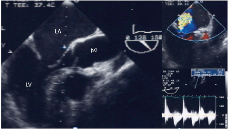
Figure 1: Multiplane transesophageal two-dimensional echocardiographic study, showing both mitral valve leaflets thickened and with reduced mobility. The interrogation with color and continuous wave Doppler demonstrated moderate regurgitation and mild mitral stenosis with area of 2.1 cm2
Her physical examination was significant for left-sided hemiplegia. Electrocardiogram showed atrial fibrillation. Cerebral angiography showed a thrombus in the right anterior cerebral artery. Ultrasound Doppler of the lower extremities showed embolic occlusion of the left femoral artery. Transesophageal echocardiogram demonstrated thrombi in the left atrium adhering to the interatrial septum (Figure 2) and also lodged in the left atrial appendage (Figure 3). The mechanical prosthetic mitral valve was structurally normal as shown in Figure 4. Anti beta 2 glycoprotein 1 antibody were strongly positive on laboratory analysis of the blood. She received intravenous heparin and was discharged home after one month of hospitalization in NYHA functional class II.
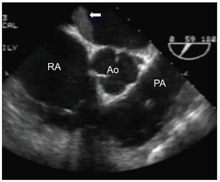
Figure 2: Transesophageal image of 4 chamber view where a thrombus adhered to the left side of interatrial septum (arrow) is observed
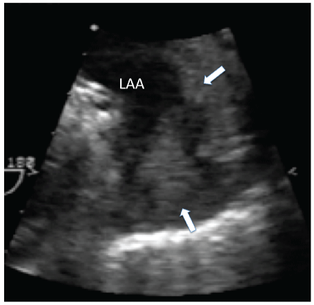
Figure 3: Thrombus in the left atrial appendage is shown (arrows)
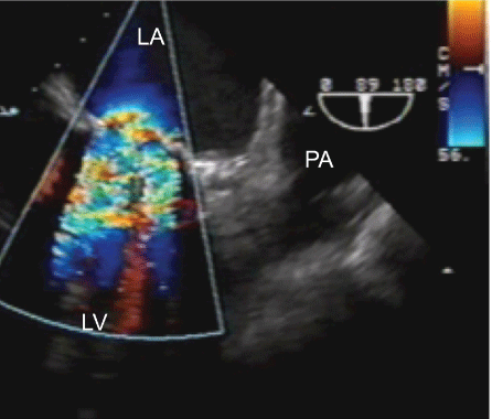
Figure 4: Bidimensional and color flow multiplane 89° transesophageal echocardiography with a mechanical prosthetic valve is showed
Discussion
The antiphospholipid syndrome is a immunothrombotic disorder characterized by the presence of antiphospholipid antibodies, leading to hypercoagulability, vascular thrombosis, and recurrent fetal loss. It usually presents in women between the ages of 20 and 40 years [1]. There are many theories that explain the prothrombotic nature, including the phenomenon of activation of the endothelial cells, binding of antiphospholipid antibodies, release of cytokines and endothelial damage by the oxidated LDL [2].
There are 3 clinically distinct forms of the disease: primary, secondary, and catastrophic. Primary antiphospholipid syndrome occurs in the absence of any underlying disorder. If the disease occurs with other autoimmune disorders, it is referred to as secondary antiphospholipid syndrome. The catastrophic antiphospholipid or Asherson syndrome is characterized by simultaneous thrombotic events occurring in multiple organ systems [3].
Cardiac involvement occurs frequently. The most common cardiac abnormality is nonbacterial thrombotic endocarditis, which is characterized by adherent platelet-fibrin thrombi on the endocardial surface of valves. In 30 to 70% of the patients the mitral valve is most frequently involved and the most common echocardiographic abnormality is a focal or diffuse leaflet thickening accompanied by reduced leaflet mobility [4]. The extent of fibrosis may determine the degree of valve thickening as well as valve retraction or mobility. Her previous transesophageal echocardiogram, before mitral valve replacement, showed that both the mitral valve leaflets were thickened with decreased mobility, but the diagnosis of antiphospholipid syndrome was only made when she presented with a stroke. The transesophageal echocardiogram showed thrombi in the left atrium as well as the left atrial appendage and the strongly positive anti beta 2 glycoprotein 1 antibodies confirmed the diagnosis.
The Asherson syndrome is fatal in approximately 50% of cases. Features of disseminated intravascular coagulation may be evident in some patients as seen in our patient. Usually, there is marked thrombocytopenia, and a Coombs positive microangiopathic-type anemia may accompany this condition [5].
Deep vein thrombosis complicated by pulmonary embolism and arterial occlusions (most commonly cerebral and peripheral) can occur with this syndrome in approximately 15 to 20% of the patients. Thrombotic microangiopathy may affect the kidneys (70%), lungs (60%), skin (60%), central nervous system (56%), heart (50%), gastrointestinal (38%) and adrenal system (26%).
Mitral and aortic valve involvement is characterized by multiple vegetations studding these leaflets coaptation surfaces. These valve lesions have a significant embolic risk up to >3 fold for cerebrovascular events.
Myocardial infarction can result from epicardial atherosclerosis or from thrombotic small-vessel disease in these patients. They can, in addition, develop segmental and global wall motion abnormalities from myocarditis and from coronary artery vasculitis.
Neurological disorders include focal cerebral or ocular thromboocclusive events [6-8]. One of the most important features is cerebral ischemia which manifests as single or multiple ischemic strokes or transient ischemic attacks [5].
Generally, the territory of the middle cerebral artery is more commonly affected.
Conclusion
This case represents an immunologic thromboocclusive disease in a woman with a mechanical prosthetic mitral valve who developed multiple embolic strokes. An immediate diagnosis and an adequate treatment played an important role in the prognosis of our patient. Although this was a case of catastrophic antiphospholipid syndrome; which is typically associated with high mortality. Early diagnosis and appropriate management lead to favorable outcomes. Treatment should be guided by a Hematologist since the patient may require intravenous corticosteroids, plasmapheresis and intravenous antibiotics in addition to the use of intravenous heparin [5].
Download Provisional PDF Here
Article Information
Article Type: Case Report
Citation: Ocaña-Arriaga VM, Waisser-Rosentein E, Chugh R, Espinola-Zavaleta N (2017) Asherson Syndrome in a Woman: The Utility of Echocardiographic Study in its Diagnosis and Treatment. J Clin Case Stu 2(6): doi http://dx.doi.org/10.16966/2471-4925.160
Copyright: © 2017 Ocaña-Arriaga VM, et al. This is an open-access article distributed under the terms of the Creative Commons Attribution License, which permits unrestricted use, distribution, and reproduction in any medium, provided the original author and source are credited.
Publication history:
Received date: 16 Nov 2017
Accepted date: 21 Dec 2017
Published date: 27 Dec 2017
