Introduction
Müllerian duct anomalies (MDAs) result from non-development or fusion of the Müllerian ducts that give rise to the uterus, fallopian tubes, cervix, and the upper two-thirds of the vagina [1]. While these anomalies are rare, with a reported incidence of between 0.5 to 5.0% in the general population [2,3], they are associated with an increased risk of infertility, endometriosis, and miscarriage [2,3]. In addition, approximately 13%– 25% of women who experience recurrent miscarriages are reported to have an MDA [2]. A proportion of MDAs, estimated to range from 30%– 50%, are also associated with renal anomalies [4,5]. This is likely to result from embryologic arrest at gestational week 8 that simultaneously affects the Müllerian and metanephric ducts [6]. Other anomalies associated with MDAs include cardiac anomalies, vertebral body anomalies, and syndromes, such as Klippel-Feil syndrome [4,7].
Uterus didelphys with hemivaginal obstruction is a rare MDA that occurs when the midline fusion of the Müllerian ducts is arrested [6,8]. It is often associated with renal agenesis ipsilateral to the obstruction, termed obstructed hemivagina and ipsilateral renal anomaly (OHVIRA) syndrome [6,8,9]. Patients with OHVIRA syndrome may present with a variety of symptoms. For example, some may present with dysmenorrhea and dysuria during adolescence [6], while others remain asymptomatic until they show fertility problems later in life [8]. In rare cases, patients may present with a huge paravaginal mass mimicking ischiorectal swelling [10] or with acute retention of urine [11]. Due to their rare and often differing presentation, the diagnosis of OHVIRA syndrome remains challenging.
Patients suspected of having an MDA are often initially referred for pelvic ultrasonography. However, magnetic resonance imaging (MRI) [6] can more accurately visualize the vaginal septum compared with ultrasound [12]. In addition, MRI does not involve ionizing radiation, allows excellent soft-tissue characterization, and permits a greater field of interrogation than ultrasonography [1,13]. Therefore, MRI is a useful, noninvasive diagnostic approach for evaluating MDAs.
We report three cases of uterus didelphys with obstructed hemivagina in young girls presenting with dysmenorrhea diagnosed by MRI.
Case 1
A 16-year-old girl presented to the emergency department with a twoweek history of lower abdominal pain. The pain was colicky in nature and more toward the right side of the abdomen, and could be relieved by analgesics. The pain was aggravated during menstruation and associated with dysuria. Menarche was at age 14 with severe primary dysmenorrhea. She had regular cycles. Her last menstrual period (LMP) was 21 days prior to presentation. She underwent laparotomy five months prior to the current presentation due to a suspected hemorrhagic ovarian cyst. No gastrointestinal tract symptoms were evident. The patient had no history of medical illness or surgical procedures and no family history of congenital anomalies. On physical examination, she had normal secondary sexual characteristics. The abdomen was soft and non-tender, with a firm pelviabdominal mass that extended up to the umbilicus.
An enhanced computed tomography scan of the abdomen and pelvis revealed the absence of the right kidney. The left kidney appeared normal, with moderate hydronephrosis. A bicornuate-like uterus was observed, with the left horn being rudimentary and smaller in size. The right endometrial cavity was distended and filled with blood or fluid. A hugely distended vagina, filled with blood or fluid, with direct continuation with the cervix was observed. Both ovaries were unremarkable; however, an irregular-shaped low density was noted lateral to the right ovary, likely representing a distended fallopian tube. No other significant abnormalities were observed. The findings were consistent with hemato- or hydrometrocolpos, and the subject was referred for MRI of the pelvis.
Pelvic MRI indicated unilateral renal agenesis of the right kidney (Figure 1a). A large cyst-like mass filled the entire pelvic cavity, extending down from the level of ischiorectal fossae up to the level of the sacroiliac joints (Figures 1b and 1c). The cyst-like mass was shown to push the uterine body upward, creating an apparent bicornuate uterus, with the left horn smaller in size (Figure 1c). Minimal free fluid could be seen in the pelvis. The pelvic MRI findings were highly suggestive of a bicornuate uterus with hematocolpos on the right side and ipsilateral renal agenesis. Based on the above findings, the subject was diagnosed with uterus didelphys associated with OHVIRA.
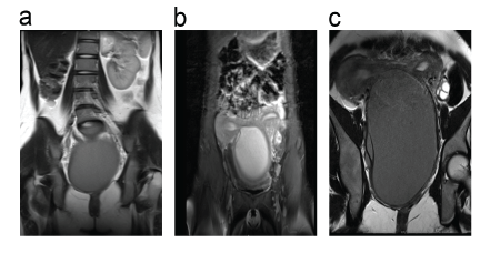
Figure 1: Selected magnetic resonance (MR, #6) images of the pelvis for Case 1.(a) Coronal T2-weighted MR image showing the absence of the right kidney with normal left kidney. A large cyst-like mass fills the entire pelvic cavity, which was relatively hypointense compared to the urinary bladder (most likely bloody content). (b) SagittalT2-weighted MR image showing the cyst-like mass pushing the uterine body upwards, with evidence of a bicornuate uterus. (c)Axial T2-weighted MR image showing that the right uterine horn is relatively larger than the left.
Case 2
A 14-year-old girl presented to the emergency department with cyclic lower abdominal pain. The pain was colicky in nature, non-radiating, and was aggravated by menstruation and movement. Menarche was at age 13. Menstruation was irregular and associated with severe dysmenorrhea. Her LMP was 5 days prior to presentation. The subject was planned for diagnostic laparoscopy and possible laparotomy in a private hospital prior to admission to our hospital. No urinary or gastrointestinal tract symptoms were evident. She had no history of medical illness or surgical procedures and no family history of congenital anomalies. On physical examination, she had normal secondary sexual characteristics. The abdomen was soft and nontender, with a soft pelvi-abdominal mass.
Pelvic ultrasonography revealed an empty right renal fossa and absence of the right kidney (images not shown. The left kidney appeared normal, without kidney stones or hydronephrosis. The uterus was retroverted and enlarged (measuring 11.1 × 5.43 × 5.96 cm), with evidence of blood within the uterus (images not shown). A partly cystic structure arose in the right iliac fossa (measuring 5.3 × 4.1 × 4.4 cm). The left ovary (measuring 3.7 × 2.1 × 3.3 cm) also contained a small cyst (measuring 2.6 × 1.5 cm). Ultrasound findings indicated potential hematocolpos, with a hemorrhagic cyst in the right ovary and right hydrosalpinx. The patient was referred for MRI of the pelvis.
Pelvic MRI revealed two myometrial cavities with two cervical canals, and a bicornuate bicornis appearance of the uterus (Figure 2). The right horn was distended with a blood product (measuring 7.1 × 6.2 × 9.8 cm in the axial, transverse, and craniocaudal dimensions, respectively). The right fallopian tube was also significantly distended (measuring 4 cm in diameter). A hemorrhagic ovarian cyst (measuring 2 × 1.8 cm) was observed on the right side. On the left side, multiple simple ovarian cysts indicative of a polycystic ovary could be seen. The left cervical canal was significantly compressed. Minimal free fluid could be seen in the pelvis.
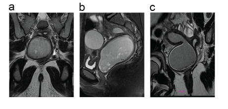
Figure 2: Selected magnetic resonance (MR, #6) images of the pelvis for Case 2. (a) TransverseT2-weighted MR image showing two myometrial cavities with two cervical canals. (b) SagittalT2-weighted MR image showing the distended right horn. (c) Axial T2-weighted MR image showing the right-sided hemorrhagic ovarian cyst.
Pelvic MR findings likely represented a bicornuate uterus with hematocolpos and hydrometrosalpinx on the right side and ipsilateral renal agenesis, as well as hemorrhagic cysts on the right ovary, a polycystic ovary on the left side, and an imperforate hymen. The subject was diagnosed with uterus didelphys associated with OHVIRA.
Case 3
An 11-year-old girl presented to the emergency department with a history of cyclic lower abdominal pain for over 1 year. The pain was colicky in nature, aggravated by menstruation, and radiated to the back. The pain was associated with constipation and dysuria, but there was no nausea or vomiting. Menarche occurred 1 year prior to presentation when the patient was 10 years old. Menstruation was irregular, minimal, and lasted for 7 days, with severe dysmenorrhea. Her LMP was 14 days prior to presentation. She had no history of medical illness or surgical procedures, and no family history of congenital anomalies. On physical examination, she had normal secondary sexual characteristics. The abdomen was soft, with slight tenderness, and a pelvi-abdominalmass extended up to the umbilicus.
Pelvic ultrasonography revealed an empty right renal fossa and absence of the right kidney (images not shown). The left kidney appeared normal, without kidney stones or hydronephrosis. The uterus was anteverted (measuring 8 × 3 × 3 cm), with endometrial thickening (8 mm). Multiple different cystic structures could be seen in the pelvic cavity, indicative of either adnexal cysts or uterine in origin. The subject was referred for MRI of the pelvis.
Pelvic MRI indicated unilateral renal agenesis of the right kidney (Figure 3a). Two endometrial cavities could be observed, one significantly distended with a blood (measuring 7.1 × 5 × 5.4 cm in the transverse, axial, and craniocaudal dimensions, respectively) and associated with hydrosalpinx (Figures 3b and 3c). Multiple cysts of varying size were observed in the right ovary (Figure 3b). The left endometrial cavity and cervical stroma were significantly stressed. The left ovary was unremarkable. Minimal free fluid could be seen in the pelvis.
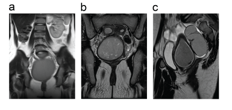
Figure 3: Selected magnetic resonance (MR, #6)images of the pelvis for Case 3. (a) Coronal T2-weighted MR image of the pelvis showing the absence of the right kidney with normal left kidney. (b) Sagittal T2-weighted MR image showing the two endometrial cavities, one significantly distended with a blood and associated with hydrosalpinx. (c) Axial T2- weighted MR image showing the right hemivagina.
Pelvic MR findings likely represented a bicornuate uterus with a rightsided hematocolpos and an imperforate hymen. The subject was diagnosed with uterus didelphys associated with OHVIRA syndrome.
Discussion
OHVIRA syndrome, formerly termed Herlyn-Werner-Wunderlich syndrome, was first reported in 1950 [14]. Onset of non-specific symptoms typically occurs during adolescence, and the mean age for diagnosis is 14 years [9]. The three cases reported here show typical presentation for OHVIRA syndrome in adolescent girls (aged 11, 14, and 16 years). In particular, the symptoms presented at puberty, with nonspecific symptoms of cyclic abdominal pain after menarche associated with a progressively enlarging pelvic mass. The secondary sexual characteristics were normal in all three cases, which were expected, as the ovarian function remains intact with this type of congenital anomaly. We used MRI to help diagnose the MDAs in these young women. In all cases, we observed a duplicated uterus. As uterus didelphys are commonly associated with renal anomalies [8], we assessed the kidneys in all three cases and found evidence of ipsilateral renal agenesis. Based on our clinical data and imaging findings, we were able to diagnose the three cases as uterus didelphys associated with OHVIRA syndrome.
MDAs must be considered as a potential diagnosis in young women presenting with a large abdominal, pelvic, or vaginal mass [10,15,16]. This is because early detection and classification of MDAs, with appropriate surgical intervention, can decrease the long-term morbidity, impaired fertility, and other obstetric complications later in life. The most widely used classification system for MDAs, which was developed by the American Fertility Society (AFS,#17) [17], includes seven subtypes: uterine agenesis/hypoplasia, unicornuate, didelphys, bicornuate, septate, arcuate, and diethylstilbestrol drug-related uterus [17].While abdominal or transvaginal ultrasound and 3D ultrasound can provide valuable information, MRI is currently the most sensitive tool for classifying MDAs according to the AFS system [1,13,18]. Magnetic resonance imaging has shown excellent agreement with the clinical diagnosis of MDA subtypes [19]. Indeed, similar to our findings, MRI was previously shown to be critical for the diagnosis and characterization of OHVIRA syndrome in previous case reports [20,21]. Therefore, our case reports further confirm the utility of MRI in the diagnosis of MDAs.
Our results also suggest that MRI should be considered as a diagnostic tool prior to laparotomy in young women presenting with dysmenorrhea and/or abdominal pain. One of the cases reported here (Case 1) underwent laparotomy 5 months prior to presentation due to a suspected hemorrhagic ovarian cyst. Another (Case 2) was planned to undergo laparotomy in the future. However, by using MRI to diagnose the MDA in the first instance, we may prevent the need for these invasive surgical diagnostic methods.
Once an accurate diagnosis is made via MRI, treatment can be tailored to the specific MDA. In terms of treatment for young women with obstructed hemivagina, resection of the vaginal septum offers symptom relief and preserves reproductive capabilities [8]. Alternatively, in cases where the obstructed hemivagina reaches the hymeneal ring, a limited resection may be performed during an initial procedure, allowing the remaining vaginal septum to be removed at a later date [22]. In addition, to preserve hymenal integrity, hysteroscopic resection of the septum under transabdominal ultrasound guidance may be performed [23]. Interventional radiology has also been used in selected cases in combination with a hysteroscopy approach [24]. Interventional radiology is also useful for directing hysteroscopic procedures or dilating occluded structures. Furthermore, in a review of 27 cases of OHVIRA syndrome over 12 years, Smith et al. found that in most patients with OHVIRA syndrome can be treated solely with single-stage vaginoplasty [9]. Moreover, they concluded that routine laparoscopy is not essential for management [9]. Therefore, MRI is useful for detecting, diagnosing, and assessing the surgically correctable forms of MDAs [25].
Conclusion
Based on our findings, we show that it is important to consider a diagnosis of MDA, including OHVIRA syndrome, when evaluating adolescents with abdominal pain and dysmenorrhea. Early diagnosis and appropriate surgical intervention in young women with these MDAs can reduce the long-term morbidity and impaired fertility. We found that MRI is a useful tool for the correct diagnosis of MDAs, and reduces the need for surgical diagnostic methods in these young women.
Acknowledgment
We thank Dr Julia Archbold, PhD, who provided medical writing services on behalf of Oxford Science Manuscripts Ltd., Oxford, United Kingdom.
Conflict of Interest
None declared.
